About telangiectasia
A telangiectasia (telangiectases or telangiectasias in plural) is a flat, dilated, red-colored blood vessel visible on the skin or a mucosal membrane, typically 0.5 mm to 1.0 mm in diameter. Related terms are: venulectasia, which refers to blue-hued vessels, somewhat raised and less than 2 mm in diameter; and reticular veins, which are 2-4 mm in diameter and raised. Most telagiectasias are not related to any other medical condition and are considered to be “primary” or “essential”. Typically, there are one or several distinct lesions, often on the face or legs.
- There are a number of medical conditions with telangiectasias as a prominant feature, including hereditary hemorrhagic telangiectasia, ataxia telangiectasia, and the CREST variant of scleroderma.
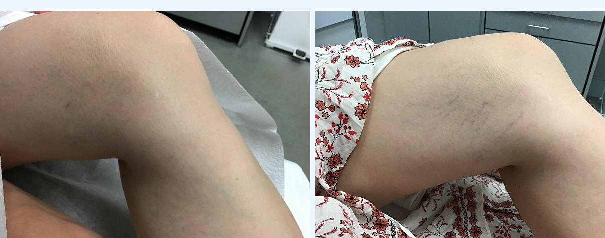
- In the legs,telangiectasias are often related to venous hypertension in the lower extremities. The co-existence of varicose veins is common. Women are more likely than men to develop these telangiectasias; pregnancy is a predisposing condition; and those who stand for prolonged periods in their daily activities are at increased risk.
- On the face, acne rosacea is often associated with the development of telangiectasias.
- Steroid treatment, both systemic and local, can cause telangiectasias.
- Telangiectasia formation following sclerotherapy is a well-known risk.
- Chronic sun and cold exposure can result in telangiectasia formation.
- Alcohol use and high estrogen levels can lead to telangiectasia, spider angiomas and palmar erythema.
Treatment for telangiectasia
The treatment for a telangiectasia depends on location, size of the vessel, how many are present and underlying etiologieis.
- Telangiectasias on the face are often treated with laser therapy with a pulsed dye laser (such as the V-Beam from Candela Laser).
- Telangiectasias and venulectasias on the lower extremities are often treated with sclerotherapy, in which a “sclerosant” in injected into the blood vessel to obliterate it. This is considered the “gold standard”.
- Blood vessels on the lower extremities are sometimes treated with laser, usually either the long-pulsed Nd:YAG or a pulsed dye laser.
- Electrocautery can be used to trace individual vessels.
- Treatment of telangiectasias on the lower extremity are at greater risk of scarring, pigmentary change or ulceration.

Before and after Vbeam for leg blood vessels. Results may vary patient to patient.
